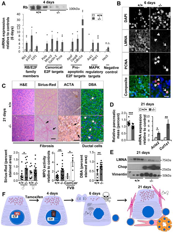
Fig. 2. LMNA-KO mice develop a phenotype that resembles chronic pancreatitis associated with RB destabilization and E2F target-activation.

(A) RB protein is decreased in LMNA-KO pancreata (4d). E2F target-transcripts were quantified using RT-qPCR (6d). N=6 mice/group. (B) Immunofluorescence of pancreata (6d) from panel-A. Solid arrows: PCNA+/LMNA- cells. Dashed arrow: PCNA+/LMNA+ cells. Bar=20μm. (C) Pancreata (21d) were stained as indicated. Arrows highlight positive staining. Bar=50μm. Histograms: Quantification of Sirius-Red, MPO-activity and DBA-staining. Wild-type mice (FVB background), injected with saline (Con) or cerulein (Cer), were used as controls for the MPO assay. (D) Relative pancreatic mass (N=13-15 mice/group) and mRNA expression of fibrosis markers determined by RT-qPCR (N=8 mice/group). (E) Immunoblot of pancreata lysates (21d) from control/KO mice (4 pancreata/genotype). (F) Proposed model of LMNA role in pancreatic homeostasis. (*p≤0.05,**p≤0.01,***p≤0.001).
