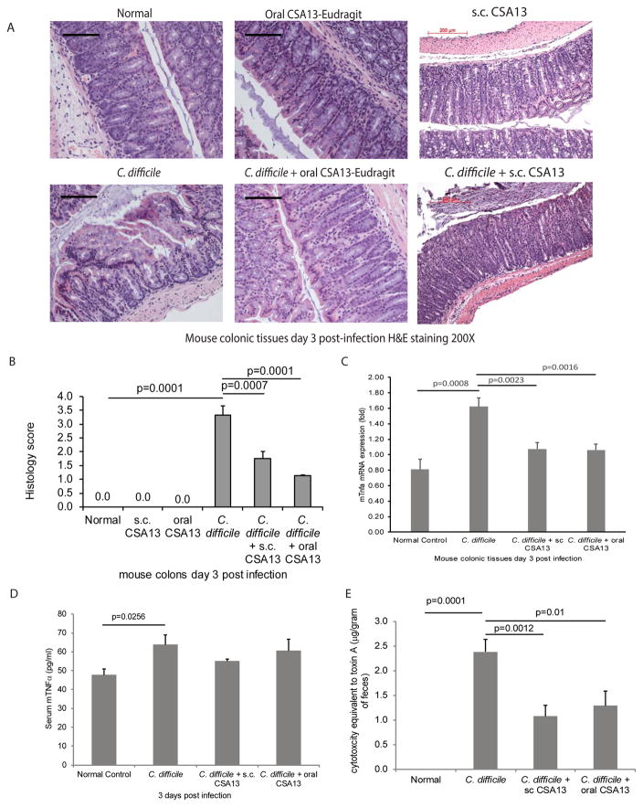
Figure 2. Subcutaneous and oral CSA13 administration reduced colonic damages in CDI.
(A) H&E staining of colonic tissues. Black bars indicate 200μm. (B) Histology score. (C) Colonic tissue TNFα mRNA expression (fold). (D) Serum TNFα levels. (E) Cytotoxicity of toxin in the sterile fecal filtrate. Each group consisted of 8 mice.

