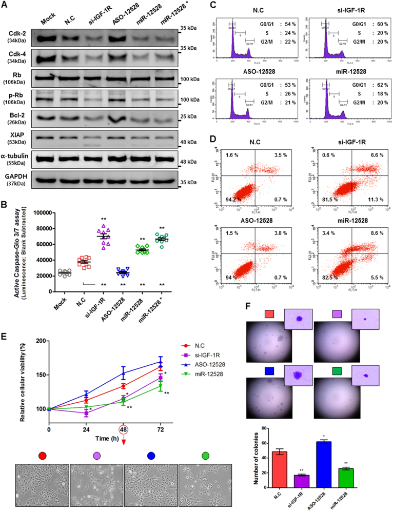
Fig. 3. The anticancer effect of hsa-miR-12528 in cell cycle and apoptosis pathway.
The signalling alteration and effect for miR-12528 on cell cycle and apoptosis pathway. Forty-eight hours post transfection, Cdk-2, Cdk-4, Rb and pRb proteins in cell cycle pathway with Bcl-2 and XIAP, anti-apoptotic proteins, were analysed using a western blotting (a). Caspase-3/-7 activity, apoptotic execution factors, assessed using a Caspase-Glo® 3/7 reagent (mimic conc.; 50 nM and *; 100 nM) (b). FACS analysis was performed using flow cytometry in A549 cells fixed or permeabilized 48 h post transfection. Cell cycle distribution was determined according to DNA stained with PI (c). Apoptotic cell death was sorted using Annexin V-FITC and PI staining. Annexin V-FITC (Annexin V+/PI−) indicates that the distribution of apoptosis in the inner leaflet of the phospholipid bilayer of the plasma membrane is translocated to the outer leaflet where the plasma membrane is intact. Annexin V+/PI+ indicates the distribution of late apoptotic cell death (d). Influence of miR-12528 on cell viability in vitro. In short-term proliferative rates, A549 cells were determined to be time-dependent via a XTT assay after transfection with 100 nM miRNA mimics. Cell morphology was imaged at 48 h (×100 magnification) (e). After mimic transfection, A549 cell-derived colonies were formed in soft agar for 3 weeks at 37 °C with 5% CO2. Graphical presentation shows the representative single colonies at high-power views (×100 magnification) and the numbers of colony-forming units (f). The experiments were performed in triplicate for independent samples (bars, mean ± S.E.M.; *p < 0.05 and **p < 0.01)

