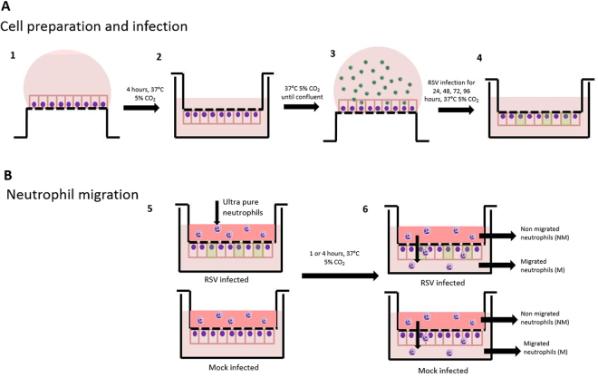
Figure 1.
Schematic diagram of neutrophil migration model. Diagram of cell seeding, A549 infection (A) and neutrophil migration (B). (1) A549 cells were seeded onto the underside of a transwell and allowed to attach for 4 hours. (2) Transwells were subsequently inverted and maintained in media to allow a confluent epithelial monolayer to develop for 72 hours. (3) Transwell were inverted and infected with GFP RSV or mock infected for 2 hours. (4) Transwells were inverted and maintained in media and infection allowed to progress for 24, 48, 72 or 96 hours. (5) Ultrapure neutrophils were added to the basolateral side of the transwell, and were allowed to migrate for 1 or 4 hours. Underneath the transwell was HBSS+ or RSV infection media. (6) Post migration basolateral and apical neutrophils were collected and downstream analysis performed.

