
Figure 4.
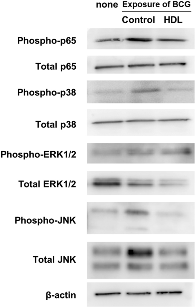
HDL impairs activation of TLR2-mediated intracellular signalings. THP1 macrophages were cultured with or without (control) adding HDL (50 µg/ml) for 24 hours. The macrophages were then infected with BCG (multiplicity of infection = 10) for 15 minutes. Immunoblot of p65 phosphorylation (phospho-p65), total p65, p38 phosphorylation (phospho-p38), total p38, ERK phosphorylation (phospho-ERK), total ERK, JNK phosphorylation (phospho-JNK) and total JNK (relative to total β-actin) were detected. Each whole cell lysate was fractionated by SDS-PAGE and transferred on a membrane.Immunoblot experiments were then performed to validate the level of each target protein. The western blot bands of phospho-p65 (65 kDa) total p65 (65 kDa), phospho-p38 (43 kDa), total p38 (43 kDa), phospho-ERK (44 and 42 kDa), total ERK (44 and 42 kDa), phospho-JNK (46 and 54 kDa) and total JNK(46 and 54 kDa) and total beta-actin (43 kDa)are displayed in the figure. Wholemembranes were also displayed in Supplemental Fig. S5. The immunoblots are representative of three independent experiments.