Figure 1.
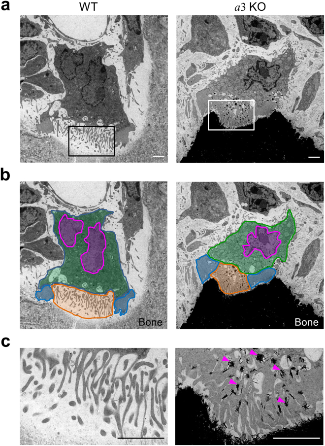
Osteoclasts in the epiphysis of humeral bones from wild-type and a3-knockout mice. Electron micrographs of a representative area containing osteoclasts in the humeral epiphysis from wild-type (a, WT) and a3-knockout (a, a3KO) mice are shown, together with coloured images (b). The ruffled border (orange), actin rings (blue), nucleus (magenta) and cytoplasm (green) are indicated schematically. An osteoclast faces the bone matrix with a finger-like ruffled border between the peripheral clear zone (actin ring). Higher magnification images of the boxed area in a are shown in c. In the mutant mouse, electron-dense material is found between processes of the ruffled border (c, arrowheads). The bone matrix is markedly more electron-opaque in the mutant mouse than in the wild-type mouse. The images are representative of 18 wild-type cells and 14 a3-knockout cells. Bars indicate 2 μm.

