Figure 2.
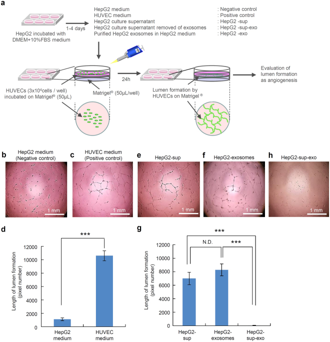
The lumen formation by HUVECs treated with HepG2-exosomes. (a) The schematic diagram of the experimental protocol is shown. The upper diagram shows the preparation of the culture medium and supplements, such as the HepG2 medium (negative control), HUVEC medium (positive control), HepG2 culture supernatant (HepG2-sup), purified HepG2 exosomes (HepG2-exosomes) and HepG2 culture supernatant with the exosomes removed (HepG2-sup-exo). The lower diagram shows the preparation used to assess the lumen formation of HUVECs in plates coated with growth factor-reduced Matrigel. The lumen formation of HUVECs on Matrigel was observed using a phase-contrast microscope. (b,c,e,f,h) The lumen formation by HUVECs under various culture conditions, such as HepG2 medium (negative control), HUVEC medium (positive control), HepG2 culture supernatant (HepG2-sup), purified HepG2 exosomes (HepG2-exosomes) added to HepG2 medium and HepG2 culture supernatant with the exosomes removed (HepG2-sup-exo). (d) The comparison of the length of the lumens formed by HUVECs between the HepG2 medium (negative control) and HUVEC medium (positive control). (g) The comparison of the length of the lumens formed by HUVECs between HepG2 culture supernatant (HepG2-sup), purified HepG2 exosomes (HepG2-exosomes) and HepG2 culture supernatant with the exosomes removed (HepG2-sup-exo). These data are shown as the means ± standard deviation of triplicate values. ***P < 0.001.

