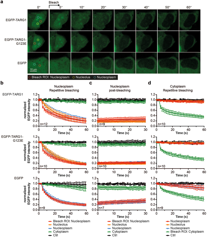
Figure 4.
EGFP-TARG1 rapidly shuttles between the nucleoplasmic and the nucleolar compartment. (a) Fluorescence loss in photo-bleaching (FLIP) experiments in U2OS cells transiently expressing EGFP-TARG1, EGFP-TARG1-G123E or EGFP. mCherry-H2B was co-expressed to define the nucleoplasm (not shown). Representative images are shown. (b–d) EGFP fluorescence intensities of EGFP-TARG1 (upper panels), EGFP-TARG1-G123E (middle panels) and EGFP (lower panels) were measured in each ROI during photo-bleaching of nucleoplasmic EGFP (b), after total photo-bleaching of nucleoplasmic EGFP (c) or during photo-bleaching of cytoplasmic EGFP (d). Fluorescence intensities were normalized to the mean EGFP fluorescence intensity in the same ROI before bleaching and are expressed as mean ± SD of 7–12 cells as indicated.

