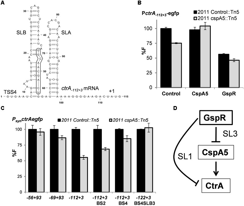
FIGURE 6.

CspA5 facilitates ctrA translation. (A) RNAfold predicted secondary structure of ctrA-112+3 mRNA transcripts exhibiting two stem loop (SL) structures. Nucleotide positions relative to the 5′-end of TSS4 are indicated. The 10-nt GspR interaction region on ctrA mRNA SLB is boxed. Means of relative fluorescence intensity values in the Rm2011 Control::Tn5 or Rm2011 cspA5::Tn5 background cells: (B) co-transformed with ctrA-112+3 translational fusion and control plasmid pSRKGm, PlaccspA5 or pSKGspR+ in the presence of IPTG; (C) transformed with the indicated ctrA native translational fusions or its mutant variants. Fluorescence measurements have been performed as described in Figure 4. (D) Schematic representation of the coherent feed forward loop circuit formed by the sRNA GspR and its ctrA and cspA5 mRNA targets, which are negatively regulated through GspR SL1 and SL3, respectively.
