
FIGURE 2.
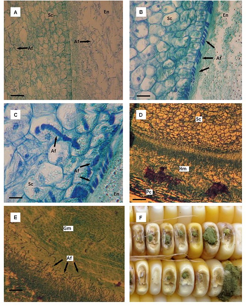
Thin tissue sections (A–E) and whole kernels (F) of aflatoxin susceptible and resistant maize hybrids colonized by Aspergillus flavus. (A) A. flavus colonized endosperm (En) and scutellum (Sc) of the susceptible hybrid 10 days after infection (DAI); starch granules in the endosperm have disappeared and are replaced by mycelium. (B) A. flavus hyphae massed at the endosperm (En)-scutellum (Sc) interface in the resistant hybrid 10 DAI; notice the outer, organized layer of cells in the scutellum. (C) A. flavus penetration of scutellum (Sc) cells in the resistant hybrid 10 DAI. (D) A. flavus hyphal mat found between the pericarp (Pc) and the aleurone layer adjacent to the scutellum (Sc) 12 DAI in the susceptible hybrid. (E) A. flavus invasion of germ (Gm) tissue of the susceptible hybrid 12 DAI. (F) Typical A. flavus growth and sporulation on needle-inoculated kernels. Scale bars: (C) 25 μm; (B) 50 μm; (D) 75 μm; (A,E) 100 μm.