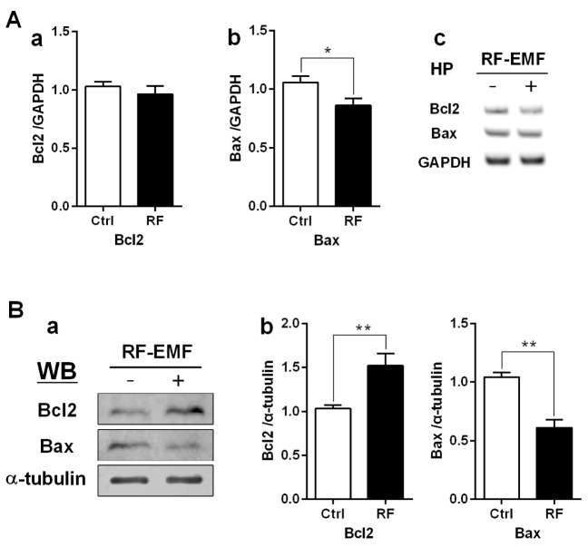
Fig. 2. Regulation of apoptosis in the mouse hippocampus following RF-EMF exposure.
Hippocampal mRNA (A) and proteins (B) were analysed for the expression level of apoptotic genes/proteins. (Aa, Ab) Quantification of Bcl2 and Bax mRNA transcripts by qRT-PCR. (Ac) Representative gel images of Bcl2 and Bax by sqRT-PCR. The expression levels of the hippocampus of RF-EMF-exposed mice were normalized to those of the sham-exposed mice. The relative mRNA levels of each gene were calculated by normalizing to GAPDH expression by the 2−ΔΔCt method (n=10). (Ba) Representative immunoblots of hippocampus from sham-control and RF-EMF group. (Bb) The expression level of Bcl2 was significantly increased, but the expression level of Bax was significantly decreased after 835 MHz RF-EMF exposure for 4 weeks. *p<0.05 and **p<0.01, vs. Ctrl. Average is the mean±SEM (n=10).

