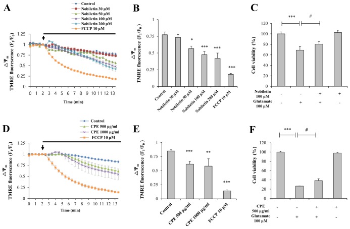
Fig. 2. The effects of nobiletin on basal ΔΨm and cell viability against glutamate toxicity in primary cortical neurons.
(A, D) Recording traces of ΔΨm using real-time imaging-based fluorometry with TMRE (see ‘METHODS’ for the detailed description). Various concentrations of nobiletin and CPE were superfused over primary cortical neurons on a cover slip in a recording chamber from the arrow point. TMRE fluorescence values from individual cells were normalized to values before drug treatment shown as an arrow. (B, E) Quantification of ΔΨm at the end of experiment for panel A and D. (C, F) Effects of nobiletin and CPE on cell viability against glutamate toxicity (100 µM, 20 min) were investigated using MTT assay. Values are the mean±S.E.M. *p<0.05, **p<0.01, ***p<0.001 as compared with the control group and #p<0.05 as compared with glutamate alone-treated group.

