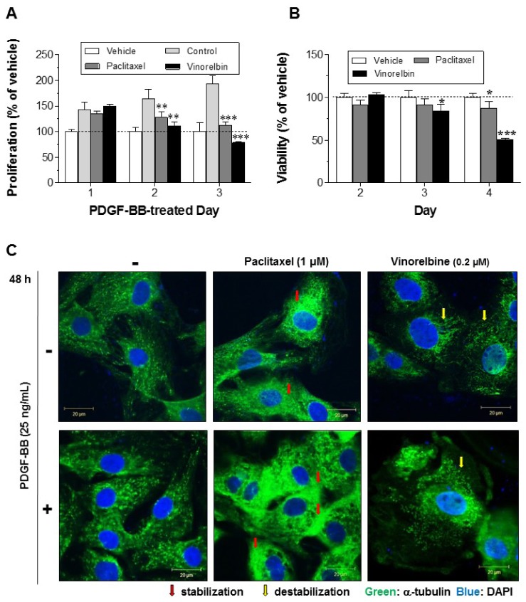
Fig. 1. Effects of paclitaxel and vinorelbine on VSMC proliferation, viability and microtubule regulation.
Serum-starved VSMCs were incubated with 1 µM paclitaxel or 0.2 µM vinorelbine for 24 h followed by 25 ng/ml PDGF-BB-treatment for 24–2 h. VSMC proliferation and viability were evaluated using the MTT assay (A, B). Mean values of the vehicle group (0.1% DMSO) were set to 100%. Data are expressed as means±SEM (n = 3). *p<0.05, **p<0.01, ***p<0.001 vs. control (PDGF-BB alone) or vehicle. (C) Microtubules were observed by confocal microscopy. After cells were fixed with 4% formaldehyde and membrane-permeabilized using 0.25% Triton X-100, immunofluorescence staining was performed using anti-α-tubulin and anti-FITC antibodies. Nuclei were stained with DAPI. Red arrow indicates stabilization of microtubule and yellow arrow indicates destabilization of microtubule. Immunofluorescence images are representative of those obtained from three independent experiments (scale bar: 20 µm; nuclei: blue; α-tubulin: green).

