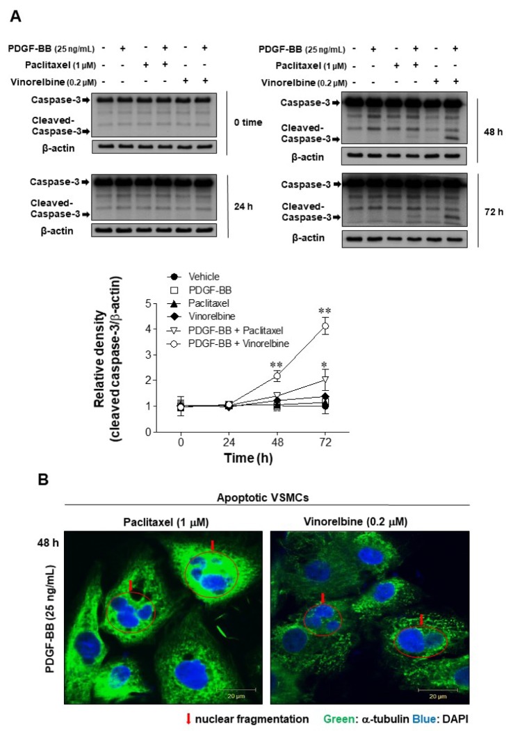
Fig. 2. Effects of microtubule regulation on caspase-3 in PDGF-BB-stimulated VSMCs.
Serum-starved VSMCs were incubated with 1 µM paclitaxel or 0.2 µM vinorelbine for 24 h followed by 25 ng/ml PDGF-BB treatment for 24–72 h. (A) VSMC apoptosis was evaluated according to the levels of caspase-3 cleavage (an apoptosis marker). The levels of full-length caspase 3 (35 kDa) and the cleaved fragment (17–19 kDa) were assessed by western blotting. The band densities were normalized to those of β-actin. The gel images shown are representative of those obtained from three independent experiments. The relative density was plotted by line graph. Mean values of the vehicle group (0.1% DMSO) were set to 1 fold. Data are expressed as means±SEM. *p<0.05, **p<0.01 vs. vehicle. (B) Immunofluorescence staining was performed using anti-α-tubulin and anti-FITC antibodies, and the VSMC nuclei were stained with DAPI (arrow: nuclear fragmentation; scale bar: 20 µm; nuclei: blue; α-tubulin: green).

