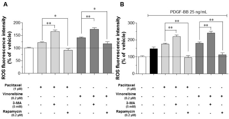
Fig. 5. Intracellular ROS levels through microtubule regulation and autophagy in PDGF-BB-stimulated VSMCs.
Serum-deprived VSMCs were incubated with 1 µM paclitaxel, 0.2 µM vinorelbine, 5 mM 3-MA (autophagy inhibitor), or 0.2 µM rapamycin (autophagy stimulator) for 24 h followed by 25 ng/ml PDGF-BB treatment for 48 h. After stimulation, the cells were stained with 20 µM H2DCFDA for 30 min at 37℃, and the fluorescence intensities were measured. (A) ROS levels of microtubule- and autophagy-regulated VSMCs. (B) ROS levels of microtubule- and autophagy-regulated, and PDGF-BB-stimulated VSMCs. Mean values of the vehicle group (0.1% DMSO) were set to 100%. Data are expressed as means±SEM (n=3). *p<0.05, **p<0.01 vs. the indicated group.

