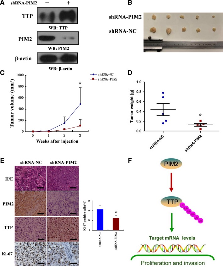
Figure 7.

PIM2‐mediated TTP function promotes breast tumor growth in vivo. (A) shRNA‐PIM2 was stably expressed in MCF‐7 cells followed by western blotting using indicated antibodies. (B) Photographs of tumors excised 3 weeks after inoculation of stably transfected cells into nude mice. (C) Tumor volumes were measured during the tumor growth for 3 weeks. Tumor volumes were calculated according to the following formula: Tumor volume = (length × width2)/2. (D) After 3 weeks, the nude mice were killed and tumor weights were measured. (E) Nude mice tumor tissues were paraffin‐embedded and tumor slides were stained with hematoxylin & eosin (H&E), antibodies of PIM2, TTP and Ki‐67 (400×). (F) Schematic diagram of the proposed PIM2 regulation of TTP degradation in breast cancer. All data are the mean ± SD of five independent experiments, *P < 0.05.
