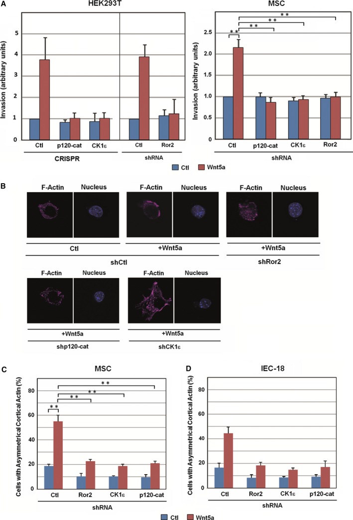
Figure 6.

The co‐receptor Ror2, p120‐catenin, and CK1ε are necessary for cell invasion and cortical actin polarization induced by noncanonical Wnt. (A) HEK293T (left) or MSC cells (right) were seeded in Transwell chambers containing 1 mg·mL −1 collagen type I. CRISPR cells or cells transfected with the indicated shRNA were used with control or Wnt5a‐conditioned medium added to the lower chamber. After 16 h (MSC) or 36 h (HEK293) of incubation, cells were fixed and stained with crystal violet, and optical density was quantified at 590 nm. Results are presented as mean ± range from two independent experiments (left), or as mean ± SD from three independent experiments (right). **P <0.01. (B) Control MSCs show a polarized cell shape at the single‐cell level in a Wnt5a‐dependent manner. Cells were transfected with the indicated shRNA and a GFP expression vector and then plated on Matrigel for 2 h with control or Wnt5a‐conditioned medium, fixed, and stained for F‐actin and nucleus (with Dapi). (C) At least 100 GFP‐positive cells were counted for each condition, and cells with polarized actin were represented as percentage of total cells. Results are presented as mean ± SD from three independent experiments. **P <0.01. (D) IEC‐18 cells were transfected with the indicated shRNA and a GFP expression vector and stained for F‐actin. The percentage of GFP‐positive cells showing cortical actin was represented as above. Results are presented as mean ± range from two independent experiments.
