
Figure 3.
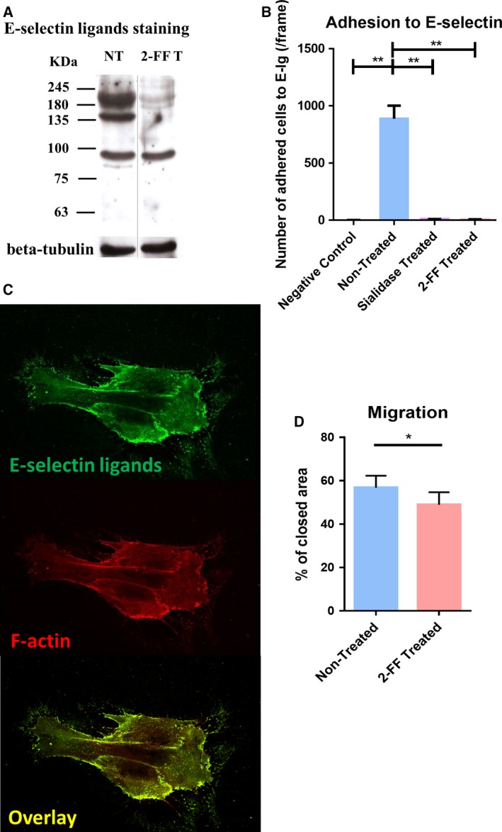
CF1_T cell line treated with 2‐FF compound loses functional E‐selectin ligands and exhibits a reduced cell migration capacity. (A) Effect of 2‐FF on the expression of sialofucosylated glycoproteins by CF1_T cells. The CF1_T cell line was treated with 1 mm of 2‐FF inhibitor (2‐FF T) or not (NT), for 5 days. Total lysate proteins were stained with the HECA‐452 mAb and analyzed by western blot. Βeta‐tubulin expression was used as loading control. (B) Effect of 2‐FF and sialidase on the capacity of CF1_T cells to adhere to E‐selectin under flow conditions. The CF1_T cells were treated or not with 2‐FF for 5 days, or sialidase for 1 h, and their capacity to adhere to E‐Ig chimera was analyzed under flow conditions by an alternative Stamper–Woodruff assay. Cells were added in calcium buffer over an E‐Ig spot and incubated with an orbital rotation at 80 r.p.m. for 30 min, at 4 °C. Assays performed in EDTA buffer were used as negative control. [n = 4; P < 0.01 (**)] (C) Cellular distribution of E‐selectin ligands in CF1_T cells. Cells were stained with E‐Ig chimera plus anti‐human IgG‐FITC (top image) and Alexa Fluor 568 phalloidin (middle image) and analyzed by confocal microscopy. Overlay of both images is displayed on the bottom. (D) The migratory ability of CF1_T cells after 10 days of 2‐FF treatment was analyzed by scratch wound‐healing assay [n = 5; P < 0.05 (*)].