Figure 6.

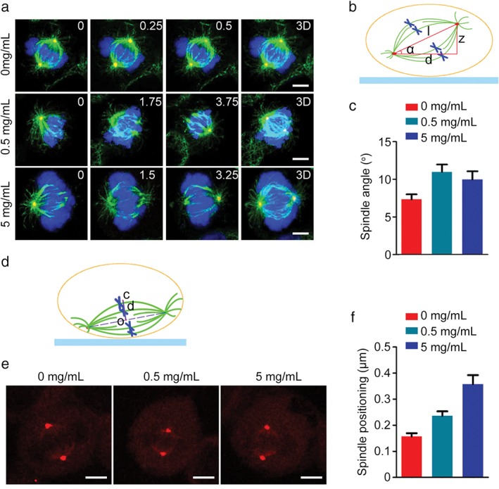
DSPS impairs spindle orientation and positioning. (a) HeLa cells treated with various concentrations of DSPS were stained with α‐tubulin (green) and γ‐tubulin (red) antibodies and 4′, 6‐diamidino‐2‐phenylindole (blue). The position of the z stage is indicated in micrometers; 3D, xy projection. Scale bars, 5 μm. (b) A diagram illustrating the spindle angle (α) measurement. (c) Determination of average spindle angle of cells treated as in panel (a). 0 mg/mL, 0.5 mg/mL, 5 mg/mL. (d) A diagram illustrating the distance (d) between the cell center (c) and the spindle center (o). (e) HeLa cells treated with DSPS were immunostained with γ‐tubulin to visualize spindle poles. Scale bars, 5 μm. (f) Quantification of the distance between the cell center and the spindle center. 0 mg/mL, 0.5 mg/mL, 5 mg/mL.
