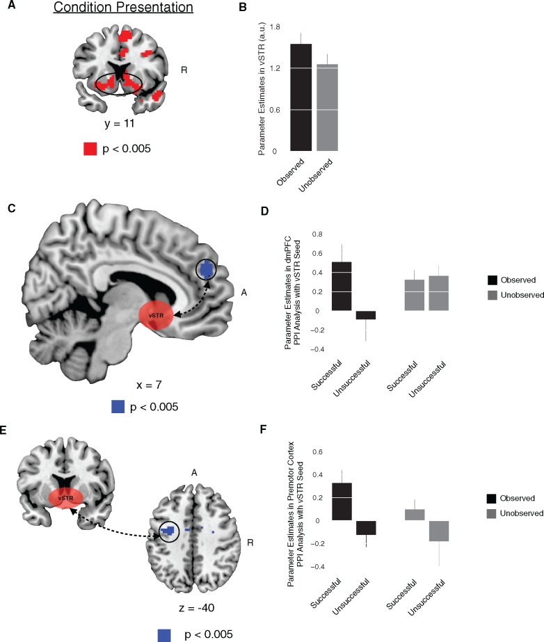
Fig. 3.
Ventral striatum and social facilitation. (A) At the time of condition presentation, vSTR exhibited significant increases in signal when comparing conditions in which participants’ performance was observed and unobserved. (B) Bar plot of the vSTR response for observed and unobserved conditions. (C) A connectivity analysis with vSTR seed, at the time of condition presentation, found that the functional coupling between vSTR and dmPFC was significantly increased during trials in which participants were observed and subsequently performed successfully. (D) Bar plot of the dmPFC PPI effects for the observed/unobserved conditions and successful/unsuccessful performance. (E) A connectivity analysis with a vSTR seed, at the time of task execution, found that the functional coupling between vSTR and premotor cortex was significantly increased during trials in which participants were observed and they were successful. (F) Bar plot of the premotor cortex PPI effects for the observed/unobserved conditions and successful/unsuccessful performance. All contrasts are significant at P < 0.05, small volume corrected. Error bars denote SEM. R, right. A, anterior.

