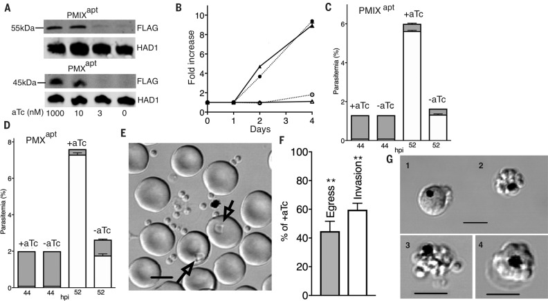
Fig. 1.
PMIX is essential for invasion of erythrocytes; PMX is essential for egress and invasion. (A) Immunoblot with aFlag antibody showing knockdown (KD) of PMIX and PMX after anhydrotetracycline (aTc) withdrawal for one cycle. Haloacid dehalogenase–like hydrolase (HAD1) was used as a loading control. (B) Expansion of PMIXapt and PMXapt parasites is impaired in –aTc medium (P < 0.0001 for each by two-tailed t test). Triangles, PMIXapt; circles, PMXapt. Open symbols, –aTc; closed symbols, +aTc. (C and D) PMIX KD results in an invasion defect. PMX KD results in both egress and invasion defects. Synchronized ring-stage cultures were grown with or without aTc. Schizonts and rings were counted by flow cytometry at 44 and 52 hours postinvasion (hpi). Shaded bars, schizonts; open bars, rings. (C) 52-hour rings were fewer in the –aTc condition [P < 0.0001 (t test)]. Number of schizonts was not significantly different. (D) 52-hour rings were fewer in the –aTc condition (P < 0.0001). Number of remaining schizonts was greater (P < 0.001). (E to G) Live-cell microscopy of PMX parasites with or without aTc. (E) Individual schizonts were scored for egress and subsequent invasion (arrows in image of control parasite field). Scale bar, 5 μm. (F) Quantification of PMXapt egress and invasion defects in the –aTc condition (**P < 0.01). (G) Abnormal schizont classes observed after PMX KD: 1, distorted schizont; 2, unruptured merozoite cluster; and 3, defective egress or merozoite dispersal. 4, Normal schizont for comparison. Scale bars, 5 μM. All experiments in this and subsequent figures were replicated at least three times. +aTc: 1 μM. Error bars indicate SEM.

