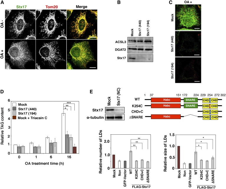
Fig. 1.
LD formation and TAG synthesis are impaired in Stx17-silenced cells. A: HeLa cells were incubated with or without 150 μM OA for 16 h, fixed, and then double immunostained for Stx17 and a mitochondrial marker, Tom20. Bars, 5 μm. B: HeLa cells were mock-transfected or transfected with siRNA Stx17 (440) or (194). After 72 h, the amounts of the indicated proteins were determined by immunoblotting. C: HeLa cells were mock-transfected or transfected with siRNA Stx17 (440) or (194). At 56 h after transfection, OA was added at a final concentration of 150 μM. After 16 h, the cells were fixed and stained with an anti-Stx17 antibody and LipidTox. Bars, 5 μm. D: HeLa cells were mock-transfected or transfected with siRNA Stx17 (440) or (194), treated with OA for the indicated times, and lysed, and then the amount of TAG was determined. As a negative control, mock-treated HeLa cells were incubated with OA in the presence of 10 μM triacsin C for 16 h, and then the amount of TAG was determined. The bar graph shows the means ± SD (n = 3). *P ≤ 0.05; **P ≤ 0.01; ***P ≤ 0.001. E: HeLa cells were mock-transfected or transfected with siRNA Stx17 (NC) targeting the 3′ noncoding region of Stx17, and the protein amounts of Stx17 and α-tubulin were determined by immunoblotting (upper left). Alternatively, HeLa cells or HeLa cells expressing the indicated FLAG-tagged constructs were transfected with siRNA Stx17 (NC), treated with OA for 16 h, fixed, and then stained with an anti-FLAG antibody and LipidTox. The bar graphs show the average number (lower left) and size (lower right) of LDs under each condition. Values are the mean ± SD (n = 3). *P ≤ 0.05; **P ≤ 0.01. “Non” denotes Stx17-silenced HeLa cells in which a vector was not transfected. Expression of an unrelated protein (GFP) had no effect on LD formation.

