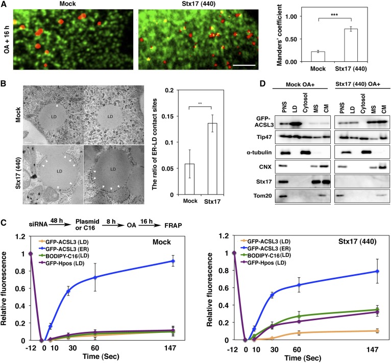
Fig. 4.
Stx17 regulates the redistribution of ACSL3 to LDs. A: HeLa cells transiently expressing GFP-KDEL were mock-transfected or transfected with siRNA Stx17 (440), treated with OA for 16 h, fixed, and then stained with LipidTox. Bar, 5 μm. The degree of colocalization between LipidTox and GFP-KDEL was measured with Manders’ coefficients. The bar graphs show the means ± SD (n = 3). ***P ≤ 0.001. B: Electron micrographs of mock-treated (upper row) and Stx17-silenced cells (lower row) after 16 h OA treatment. Bars, 200 nm. The graph on the right shows the quantitation of EM data. The ratio of the area of ER-LD contact sites relative to the circumference of the LD surface was plotted. Values are the means ± SD (n = 5). **P ≤ 0.01. More than 30 LDs were analyzed in each experiment. C: HeLa cells were treated as depicted in the figure, and then FRAP experiments were performed. The graphs show the relative signal intensity after photobleaching. Values are the means ± SD (n = 3). D: Mock-treated and Stx17-silenced HeLa cells transiently expressing GFP-ACSL3 were incubated with OA for 16 h, and then subjected to LD fractionation analysis. PNS, postnuclear supernatant; MS, microsomes; CM, crude mitochondria.

