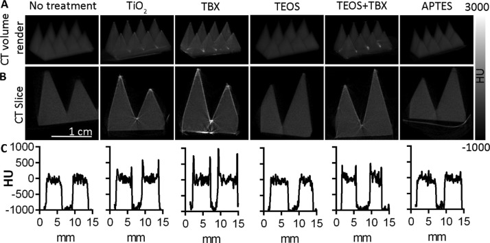
Figure 6.
CT images of treated and untreated 3D printed scaffolds, with grayscale values corresponding to their radiopacity in Hounsfield units (HU). (A) Three-dimensional volume rendered CT image showing increased surface radiopacity following treatment with TiO2, titanium(IV) butoxide, and TEOS–titanium(IV) butoxide coatings. TEOS and APTES-treated scaffolds show a radiopacity similar to that of the untreated material. (B) CT cross sections show that the chemical treatments are limited to the surface in the cases of TiO2, titanium(IV) butoxide, and TEOS–titanium(IV) butoxide, whereas TEOS and APTES are not detectable because of their radiopacity comparable to that of the untreated scaffolds. (C) CT signal intensity (HU) along a line horizontally bisecting the corresponding scaffolds shown above in (A,B).

