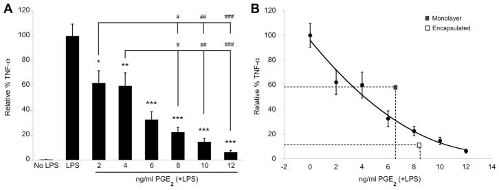
Figure 4.
(A) Rat TNF-α ELISA of cell culture media supernatant collected from organotypic cultures after 24 h of LPS stimulation ± human PGE2. Data are normalized to untreated LPS-stimulated OHSC and represented as mean ± SE from three experiments, each with n = 3 cultures per condition. Addition of exogenous human PGE2 significantly reduced TNF-α levels in a dose-dependent manner. *P < 0.01, **P < 0.005, ***P < 0.0001 compared with LPS + no treatment. #P < 0.01, ##P < 0.001, ###P < 0.0001 between dose groups. (B) Polynomial curve fit of data presented in A, overlaid with corresponding mean levels of PGE2 production and TNF-α reduction by monolayer and encapsulated MSCs.

