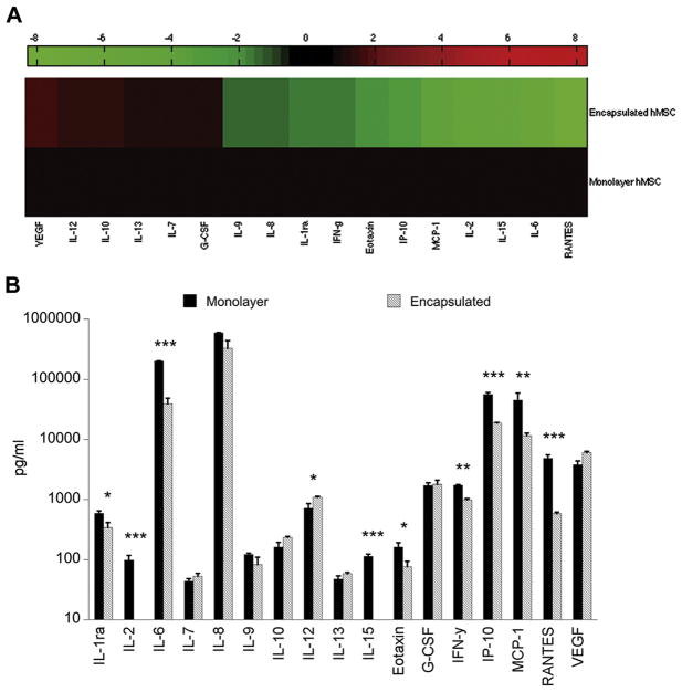
Figure 5.
(A) Heat map representation of multiplex (human) secretome data. Secretion by encapsulated MSCs (1 × 10⁵ cells/well) co-cultured with LPS-stimulated OHSC is normalized to secretion by monolayer MSCs (1 × 10⁵ cells/well) co-cultured with LPS-stimulated OHSC. Increased levels of secretion are represented in shades of red and decreased levels in shades of green. (B) Multiplex analysis of cell culture media collected after 24 h of MSC co-culture with LPS-stimulated hippocampal slices. Data are represented as mean ± SE from one experiment, with n = 3 cultures per condition. Of 17 detectable analytes, 10 were identified as exhibiting significantly different levels of secretion by encapsulated MSCs as compared with monolayer MSCs. *P < 0.05, **P < 0.005, ***P < 0.0001.

