
Fig. 2.
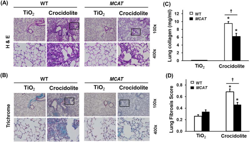
Compared to WT mice, MCAT mice are protected from pulmonary fibrosis after asbestos exposure. WT and MCAT Mice were treated with a single intra-tracheal (IT)-instillation of 100 μg in 50 μL PBS titanium dioxide (TiO2, an inert particle) or 100 μg crocidolite asbestos in 50 μL PBS and three weeks later the lungs were harvested as described in the Material and Methods and subjected to hematoxylin and eosin (H & E, A) and trichrome (B) staining, lung collagen levels (C) and lung fibrosis scores (D) as described in the Material and Methods. (A and B) Shown is representative histology from 4 to 9 mice in each group. Upper row, scale bar=0.05 mm; lower row scale bar=0.1 mm. Squares on the upper rows were enlarged in the respective lower rows. TiO2-WT or MCAT (n=4–6); crocidolite-WT or MCAT (n=7–9). The Fibrosis score =(severity: 0–4)×(extent: 1–3). (C) Lung fibrosis scores. *p < 0.05 vs. TiO2, †p < 0.001 vs. WT+crocidolite asbestos, n =4–6. (D) Collagen levels. *p < 0.05 vs. TiO2, †p < 0.05 vs. WT+crocidolite asbestos. n=7–9.