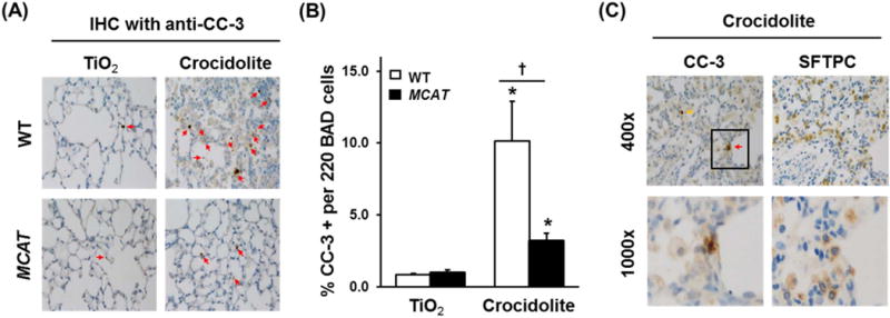
Fig. 4.

Asbestos-induced apoptosis (CC-3 activation) in cells at the BAD junction is reduced in MCAT mice versus WT mice and co-localization of CC-3- and SFTPC-(AT2)-positive cells in asbestos-exposed mice. Lungs from WT and MCAT mice were harvested 3 weeks after the IT-instillation of TiO2 or crocidolite asbestos and serial sections were subjected to IHC for CC-3 (marker of apoptosis) or SFTPC (marker for AT2 cells) IHC as detailed in the Material and Methods. (A) Apoptosis as assessed by anti CC-3 IHC in WT mice (top row) and MCAT (bottom row). (B) Semi-quantitative analysis of 220 cells at the BAD junctions. *p < 0.05 vs. TiO2, †p < 0.001 vs. WT+crocidolite asbestos, n =4–6. (C) IHC for CC-3 and SFTPC was performed on serial lung sections. Red arrows mark CC-3 and SFTPC positive cells (apoptotic AT2 cell). Yellow arrows mark CC-3 positive, SFTPC negative cells (apoptotic non-AT2 cells). Squares on the upper rows were enlarged in the respective lower rows.
