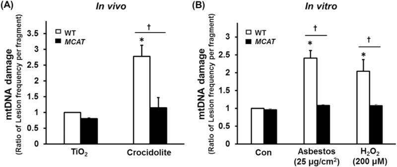
Fig. 5.

Compared to WT, AT2 cell from MCAT mice have reduced mtDNA damage after asbestos exposure in vivo (A) and in vitro (B). (A) Primary AT2 cells were isolated from the lungs of WT and MCAT mice three weeks after IT instillation of TiO2 or crocidolite asbestos and mtDNA damage were assessed by a fluorescent-based PCR mtDNA damage assay as described in the Material and Methods (expressed as the ratio of lesion frequency per fragment as compared to WT AT2 cells exposed to control – TiO2). *p < 0.05 vs. TiO2, †P < 0.05 vs. WT +crocidolite asbestos, n=4. (B) Primary AT2 cells were isolated from WT and MCAT mice, then treated with amosite asbestos or H2O2 in vitro and mtDNA damage was assessed as above (expressed as the ratio of lesion frequency per fragment as compared to WT AT2 cells exposed to control media). *p < 0.05 vs. Control of WT, †p < 0.05 vs. WT, n=4.
