
Fig. 7.
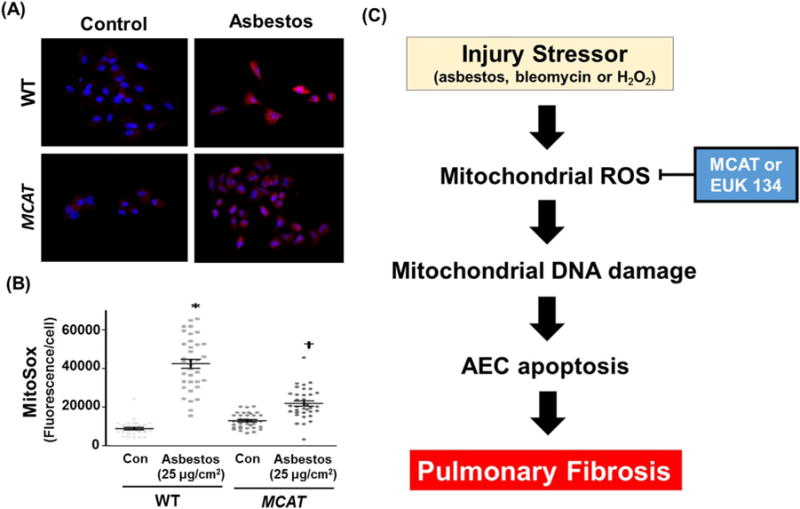
Asbestos-induced mitochondrial ROS production is reduced in AT2 cells from MCAT as compared to WT mice. (A) Primary AT2 cells were isolated from WT and MCAT mice and exposed to control media or amosite asbestos (25 μg/cm2). After 24 h, the AT2 cells were incubated with MitoSox red (90 nM) according to the manufacturer’s recommendation; shown is a representative panel from a total of 3 experiments. White punctate cytosolic staining indicative of mitochondrial ROS production as well as DAPI-stained nuclei were assessed. (B) Semi-quantitative analysis of 3 separate AT2 cell isolations from WT and MCAT mice. The graph depicts the MitoSox fluorescence intensity (mean ± SEM) in WT and MCAT murine AT2 cells; each point represents individual AT2 cell fluorescence intensities from 10 to 15 cells per experiment, lines represent mean fluorescence and brackets show standard error. *p < 0.05 vs. control/WT. †p < 0.05 vs. control/MCAT. (C) A proposed model showing that strategies aimed at reducing AEC mitochondrial ROS production (i.e. mitochondrial catalase [MCAT] or Euk-134) will attenuate AEC mitochondrial dysfunction, mtDNA damage, and apoptosis important for promoting lung fibrosis.