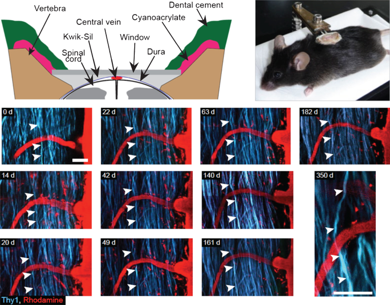
Fig.3.
Spinal glass window for long term recurrent imaging of the spinal cord. Upper panel (right) Detailed view of spinal glass window. The edges of the vertebrae are shaped so that the window rests on both sides of the vertebral opening without compressing the spinal cord. Kwik-Sil is used as a physical barrier preventing the infiltration of opaque tissue between the window and spinal cord. Upper panel (left) Image of an anaesthetized mouse with an implanted window. The window structure is being supported by a clamp attached to a plastic base. The animal is freely breathing while in the support, but the window remains stationary relative to the clamp. Lower panel: Long-term spinal cord integrity after glass window implantation. Recurrent imaging sessions (from day 0 to day 350) of the same region of a Thy1-CFP mouse spinal cord after using Rhodamine– Dextran to visualize the vascular compartment. Note that individual axons can be followed for approximately one year. Scale bar, 50 μm; Excitation wavelength 945 nm (adapted from Ref. 27).

