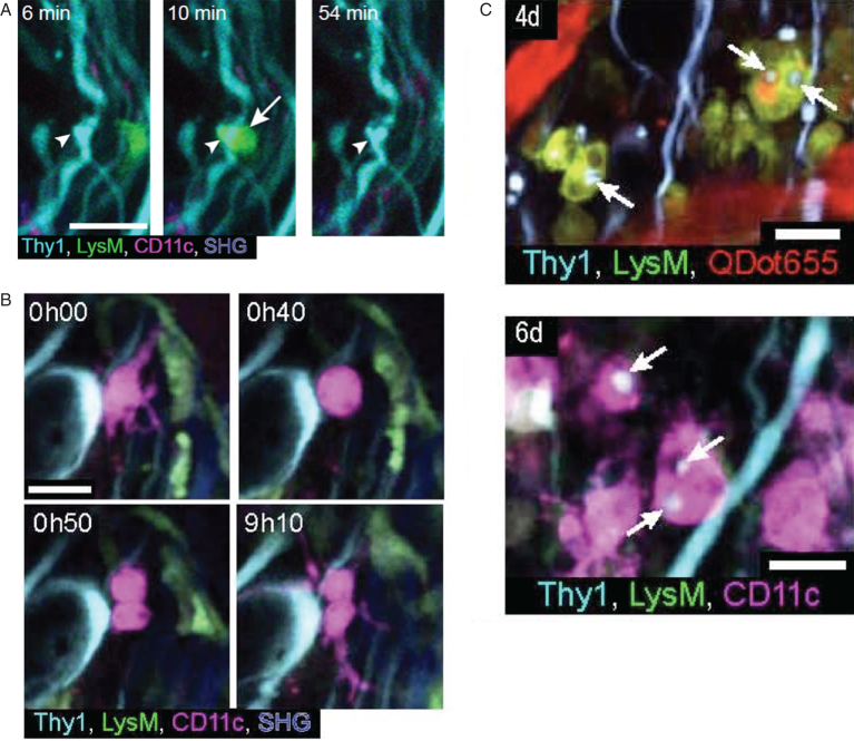
Fig.4.
Time series showing cellular events occurring after spinal cord injury: (A) time-series images (60 min) of a LysM positive cell (arrow) that formed a close apposition with an injured axon with a tortuous morphology and irregular swellings (arrowhead). (B) time-series (9 h) images of a CD11c positive cell undergoing cell division at 3 days post-injury. (C) two-photon image stack near a lesion site showing LysM positive cells and CD11c positive cells with amoeboid morphology and containing CFP positive vacuoles (arrows) at 4 and 6 days post-injury, respectively. Thy1-CFP dorsal column axons, LysM-GFP recruited peripheral myeloid cells, CD11c-EYFP microglial cells, QDot655 in blood vessels and second harmonic generation (SHG) from collagen in dura mater. Scale bars: 20 μm. (adapted from Ref. 55).

