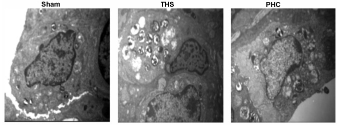
Figure 5.
Detection of lung injury by transmission electron microscopy (original magnification, ×10,000). In the sham group, the edge of the nuclear membrane was clear. The THS group exhibited significant pulmonary injury (degranulation of pulmonary alveolar type II cells, emptied osmiophilic lamellar bodies and obscured or disappeared cellular ridge). In the PHC group, a comparatively greater number of lamellar bodies was evident. THS, blunt chest trauma with hemorrhagic shock; PHC, penehyclidine hydrochloride.

