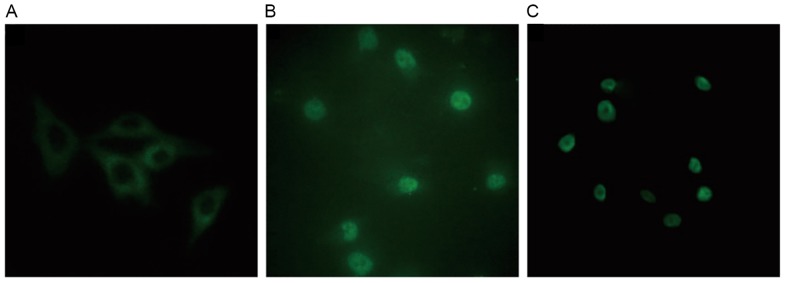
Figure 9.
Immunocytofluorescent analysis of PKC-β1 localization in human mesangial cells under various treatment conditions. PKC-β1 was detected by immunocytofluorescence and visualized by confocal microscopy. Magnification, ×400. Group A, control; Group B, HG+LPC; Group C, HG+LPC+atorvastatin. PKC-β1, protein kinase C-β1; HG, high glucose; LPC, lysophosphatidylcholine.

