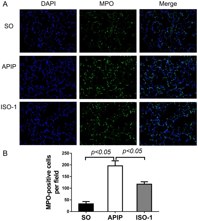
Figure 5.
Representative immunofluorescence staining for MPO of the lung sections of each group (original magnification, ×200). (A) MPO was stained green and nuclei was stained blue. (B) Comparison of the number of MPO-positive cells in the lung. P<0.05 indicates a significant difference between the marked groups. SO, sham operation group; APIP, acute pancreatitis in pregnancy group; ISO-1, ISO-1+APIP group. ISO-1, (S,R)3-(4-hydroxyphenyl)-4,5-dihydro-5-isoxazole acetic acid methyl ester; MPO, myeloperoxidase.

