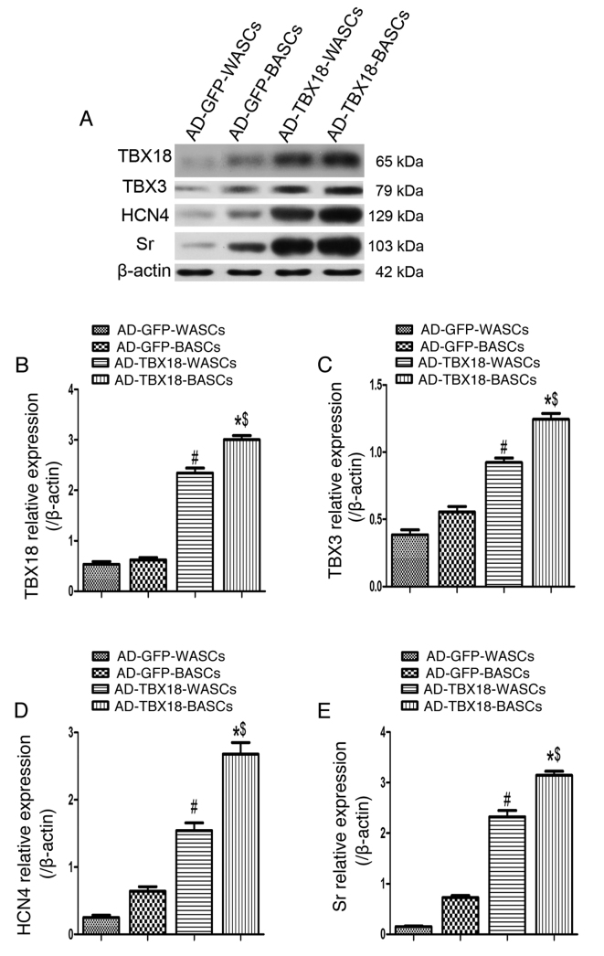
Figure 7.
Western blot analysis of pacemaker cell-associated protein expression in BASCs and WASCs following TBX18 transduction. (A) Representative western blot bands for TBX18, TBX3, HCN4 and Sr in WASCs and BASCs transduced with either AD-TBX18 or AD-GFP. Densitometric analysis was performed to quantify the protein expression of (B) TBX18, (C) TBX3, (D) HCN4 and (E) Sr. Expression levels were normalized to β-actin. Data are presented as the mean ± standard error of the mean n=3. *P<0.05 vs. AD-GFP-BASCs; #P<0.05 vs. AD-GFP-WASCs; $P<0.05 vs. AD-TBX18-WASCs. BASCs, brown adipose-derived stem cells; WASCs, white adipose-derived stem cells; TBX, T-box; HCN4, hyperpolarization-activated cyclic nucleotide-gated channel 4; Sr, sarcomeric α-actinin; AD, adenovirus; GFP, green fluorescent protein.

