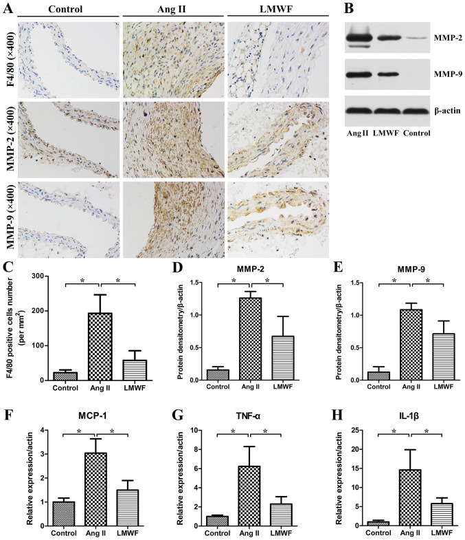
Figure 5.
(A) Representative images of immune-histochemical stains for F4/80, MMP-2 and MMP-9. Original magnification, ×400; (B) Western blot results of MMP-2 and MMP-9 in abdominal arteries; (C) Quantification of macrophage infiltration, shown as the number of positive cells per square millimeter (*P<0.05). Quantitative analysis of MMP-2 (D) and MMP-9 (E) expression (*P<0.05). mRNA expression of MCP-1 (F), TNF-α (G) and IL-1β (H) (*P<0.05). MMP, metalloproteinases; MCP, monocyte chemotactic protein.

