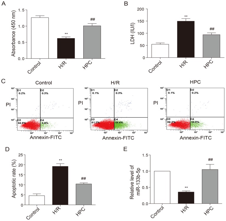
Figure 2.
Effects of HPC on H/R injury and the level of miR-133b-5p. (A) The viability of cardiomyocytes was measured by Cell Counting Kit-8 method. (B) Cell injury was determined by lactate dehydrogenase release in the culture medium. (C) Cell apoptosis was measured by Annexin V-FITC/PI flow cytometry and (D) the apoptotic rate is expressed as the percentage of cells at early apoptotic stage (the lower right quadrant). (E) The level of miR-133b-5p was detected by quantitative RT-PCR. Data are presented as mean ± SEM (n=3), **P<0.01 vs. control; ##P<0.01 vs. H/R. HPC, hypoxic preconditioning; H/R, hypoxia/reoxygenation; miR, microRNA; FITC, fluorescein isothiocyanate; PI, prodium iodide.

