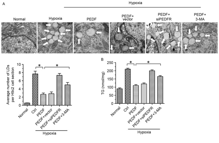
Figure 2.
PEDF-induced autophagy contributes to lipid degradation. H9c2 cells were maintained in normoxic or hypoxic conditions for 4 h with or without PEDF (10 nM). RNA interference assays were used to silence PEDF-R expression and autophagy inhibitor 3-MA (5 mM) was added. (A) Representative images of transmission electron microscopy analysis of H9c2 cells LDs. Typical LDs (arrows) were observed. Scale bar, 1 µm. (B) TG levels were quantified with a TG assay kit. *P<0.05. Ctrl, control; LDs, lipid droplets; PEDF-R, pigment epithelial-derived factor receptor; 3-MA, 3-methyladenine; siPEDF, short interfering PEDF RNA; TG, triglyceride.

