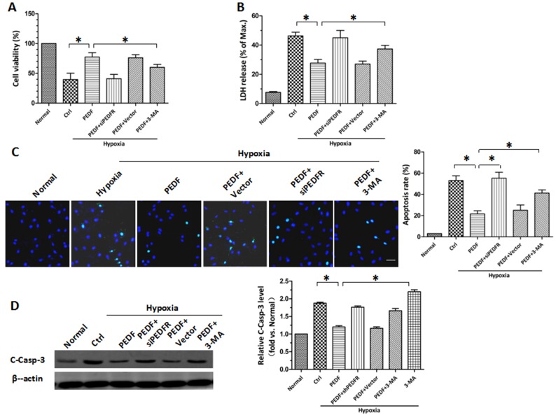
Figure 3.
PEDF protects H9c2 cells against hypoxia-induced apoptosis by inducing autophagy. CCK-8 and LDH release assays were employed to assess (A) cell viability and (B) the rate of cell death. (C) Effect of PEDF on H9c2 cells apoptosis. TUNEL (green) staining was performed within each group. Nuclei were stained with DAPI (blue). Scale bar, 50 µm. (D) Samples were collected and the expression of C-Casp-3 protein was analyzed by western blotting. *P<0.05. 3-MA, 3-methyladenine; C-Casp-3, cleaved caspase-3; CCK-8, Cell Counting Kit-8; Ctrl, control; LDH, lactate dehydrogenase; PEDF-R, pigment epithelial-derived factor receptor; siPEDF, short interfering PEDF RNA.

