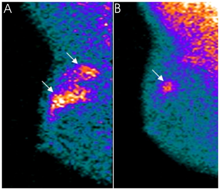
Figure 3.
A 47-year-old patient with locally advanced invasive ductal carcinoma (clinical stage at baseline: IIA) located in the external upper quadrant of the right breast clearly evident (arrows) in MBI in a the mediolateral oblique view in the (A) pre-therapy study. The patient was classified as a partial responder after neoadjuvant chemotherapy. At surgery, a unifocal residual tumor 1.5-cm large was ascertained in a (B) post-therapy MBI scan as a focal area of increased uptake (arrow) whose size corresponded exactly to the histopathological result. MBI, molecular breast imaging.

