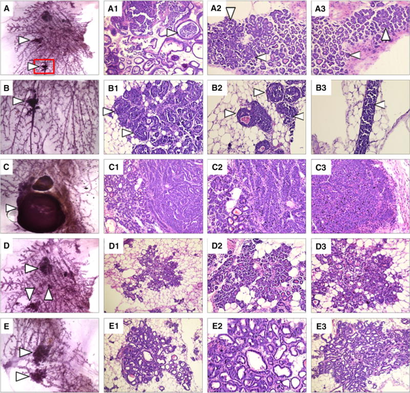
Fig. 3.

Mammary gland hyperplasia and dysplasia in NSD3 transgenic females. a Carmine staining of a whole mount of a right thoracic mammary gland harvested from an NSD3 female showing numerous areas of hyperplasia and dysplasia. A1–A3. H&E staining of sections cut from the whole mount shown in panel A detailing cystic lesions and ductal carcinoma in situ (DCIS). b Higher magnification of the area boxed in red from panel A. B1-3. Higher magnification images of H&E staining of the area shown in panel B. c Carmine-stained whole mount of the right inguinal mammary gland taken from an NSD3 female displaying a small mammary carcinoma. C1-3. H&E staining of the whole mount shown in panel C. d Carmine-stained whole mount of a right thoracic mammary gland harvested from an NSD3 female displaying numerous hyperplastic lesions. D1-3. H&E staining of the whole mount shown in panel D. e Carmine-stained whole mount of a left thoracic mammary gland harvested from an NSD3 transgenic female. E1-3. H&E staining of the whole mount shown in panel E
