Fig. 4.

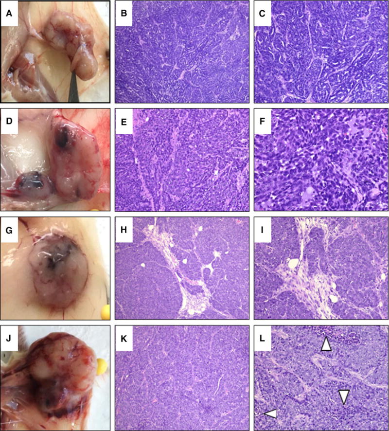
Mammary hyperplasia, dysplasia, and tumorigenesis in NSD3 transgenic females. a Tumor isolated from the left inguinal mammary gland of an NSD3 transgenic female. b–c H&E staining of histological sections of the tumor shown in panel A displaying a trabecular growth pattern. d Tumor isolated from the left inguinal gland of an NSD3 transgenic female. e–f H&E staining of histological sections of the tumor shown in panel D. g Tumor isolated from the right thoracic gland of an NSD3 transgenic female. h–i H&E staining of histological sections of the tumor shown in panel G. H&E staining shows precursor trabecular growth as well as invasive ductal carcinoma. j Tumor isolated from the cervical gland of an NSD3 transgenic female. k–l H&E staining of the tumor shown in panel J, showing invasive ductal carcinoma with trabecular growth patterns coupled with areas of necrosis
