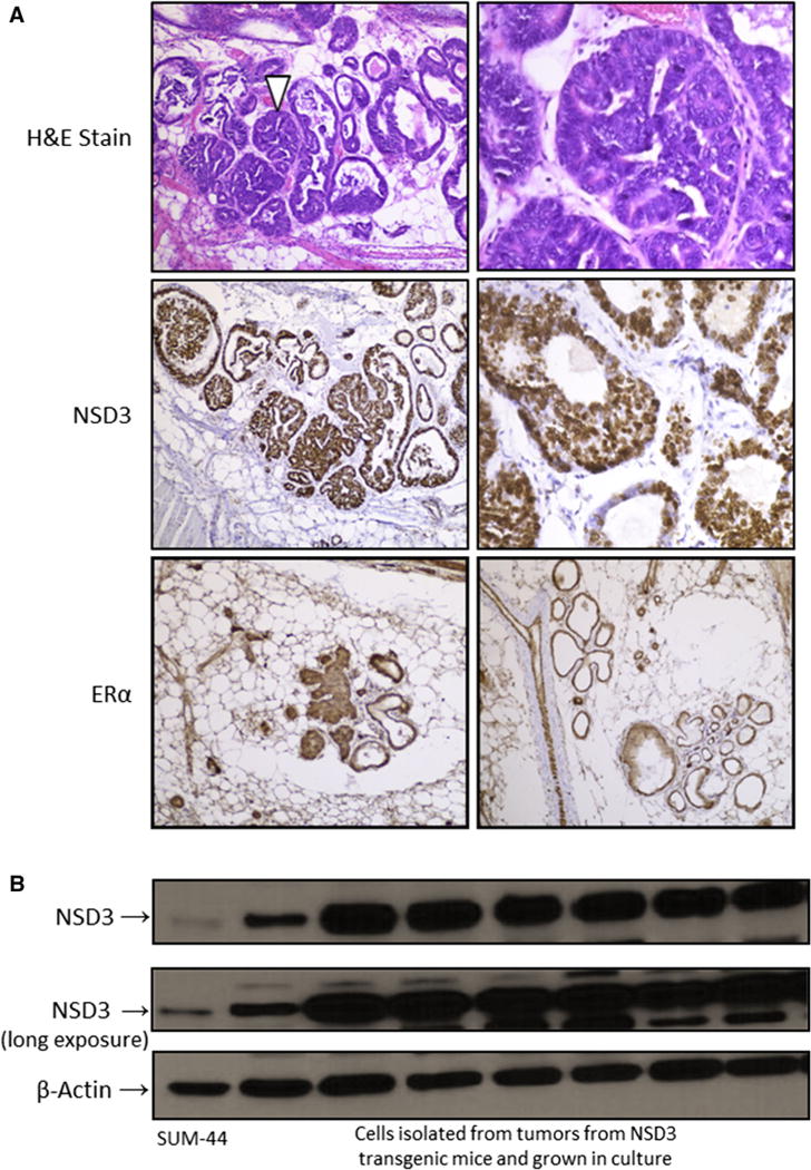
Fig. 5.

Overexpression of NSD3 in mammary tumors from transgenic females. a The upper two panels show H&E stained mammary glands from an NSD3 transgenic female exhibiting areas of hyperplasia and dysplasia. The middle two panels display positive nuclear staining for NSD3 by immunohistochemistry (IHC) in the mammary gland of an NSD3 female. The bottom two panels show positive ERa staining by IHC in mammary glands from a tumor-bearing NSD3 transgenic female. b. Western blot of NSD3 expression in cells isolated from tumors in NSD3 females and grown in culture under various conditions. Because they have been shown to express significant levels of NSD3, whole cell lysate from SUM-44 are included for comparison [16]
