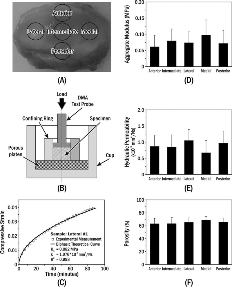
FIGURE 1.
(A) Specimen preparation. (B) Uniaxial confined compression chamber, where the polished stainless-steel confining ring prevented radial deformation and rigid permeable porous platen (20 µm average pore size) allowed water flux. (C) Typical creep behaviour of porcine TMJ disc specimen compared to biphasic theory.16 Means±standard deviations of (D) aggregate modulus, (E) hydraulic permeability and (F) porosity for specimens from five disc regions

