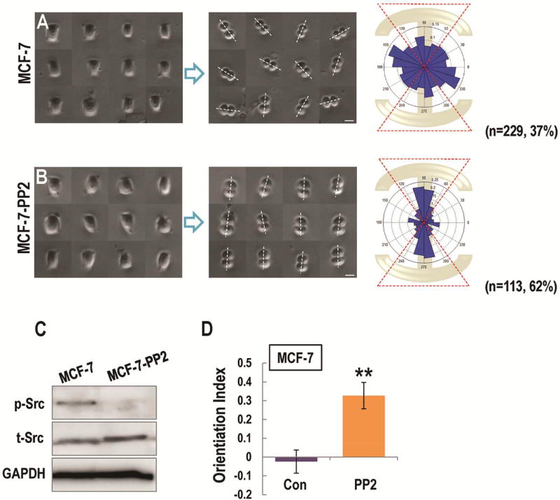
Figure 2. Src inhibition contributed to the correct orientation of mitotic cancer cells in vitro.
(A, B) Left column shows the interphase of MCF-7 (A, MCF-7), and PP2- treated MCF-7 cells (B, MCF-7-PP2) constrained on the I-type micropattern. The middle column shows the division directions in MCF-7 (A), and PP2- treated MCF-7 cells (B) after plated on I-type micropattern. The right column shows angular distribution of division orientations in MCF-7 (A), and PP2-treated MCF-7 (B) cells.
(C) The expression of total Src and phosphorylated Src in MCF-7 and PP2- treated MCF-7 cells were presented in Fig. C.
(D) Src inhibition significantly contributed to the response of cell division orientation to the geometry of micropatterns in cancerous cells. Scale bars represent 20 µm. Results represent mean ± S.D. **P <0.01 All results are representative of 3 independent experiments.

