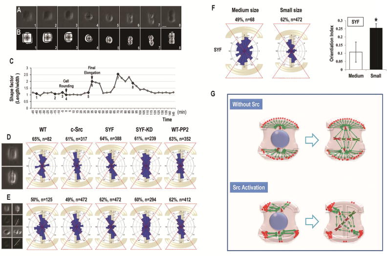
Figure 5. Src controls the accumulation and local density of cell adhesions at the site of cell matrix attachment to manipulate cell division orientation.
(A) The mitotic divisions with cell-matrix attachment on all of the four corners in SYF cells. Frames were extracted from a time-lapse phase-contrast microscopy at a rate of one picture every 5 minutes with a 10× objective. Numbers corresponded to those presented on the time curve in C. The mitotic cell center was indicated in frame 4 and the division axis was shown in frame 6.
(B) Canny edge detection to show the changes of center positions in A. Black dashed lines corresponding to the length and width of the mitotic cells were calculated on segmented pictures to show the cell shape factor. The cytoplasm in SYF cells contracted equably and finally formed a cubic shape before rounding up.
(C) Cell shape factor (ratio of length to width) shows the shape changes during mitotic division in SYF cells with four-corner attachment. Time 0 corresponded to the beginning of cell rounding.
(D) The angular distribution of cell division in WT-MEF (WT), c-Src, SYF, SYF-KD, and PP2-treated WT-MEF (WT-PP2) cells with four-corner attachment.
(E) When combining all the cell-division cases (four-corner attachment, three-corner attachment, two-corner attachment, and two opposite-corner attachment) together, the orientation of mitotic divisions in SYF, SYF-KD and PP2-treated WT-MEF cells were still oriented parallel to the long axis of the underlying micropattern, whereas the distribution of cell division axes in WT-MEF and c-Src overexpressing cells tended to be less oriented.
(F) Larger micropatterning induced the localized deprivation of cell-matrix attachment on the cell cortex and broke the balanced four-corner adhesion pattern in SYF cells.
(G) Schematic to show how Src controls the distribution of cell-matrix adhesion on the cell cortex. Without activation of Src, the cortical cue-related protein distributed homogeneously at the sites of cell-matrix adhesion, which aligned the cell division machinery along the longest axis of the cell geometry. Src activation affected the distribution of cortical cues on the cell cortex to manipulate the accumulation and local density of adhesive proteins to the sites of cell-matrix attachment, which ultimately interrupted the force balance on the spindle pole and induced the cell division misorientation. Scale bars represent 20µm. Results represent mean ± S.D. *P <0.05 All results are representative of 3 independent experiments.

