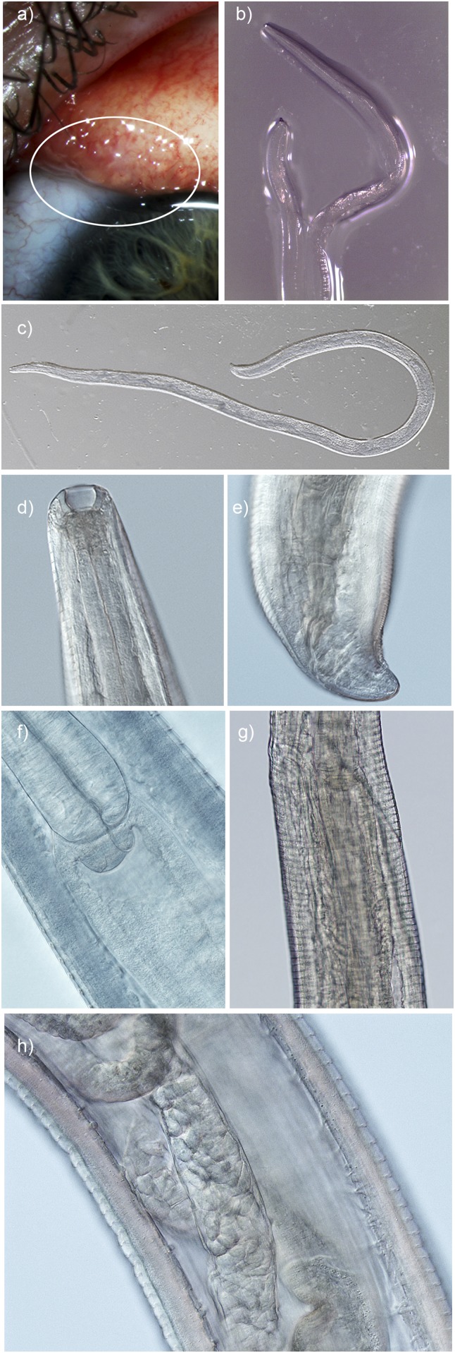
Figure 1.
Thelazia gulosa (A) in situ on the surface of the patient’s conjunctiva (circle); (B) adult female immediately after removal from the eye. Morphological identifying features of adult female worm submitted for analysis; (C) whole adult female (×40 magnification, cleared); (D) deep buccal cavity; (E) tail with nonprotruding anal opening and postanal papilla; (F) esophageal-intestinal junction; (G) nonprotruding vulval opening slightly anterior and to the left of the esophageal-intestinal junction; and (H) mid body with prominent cuticular striations, intestinal tube, and ovaries containing spirurid eggs (×200 magnification, cleared).

