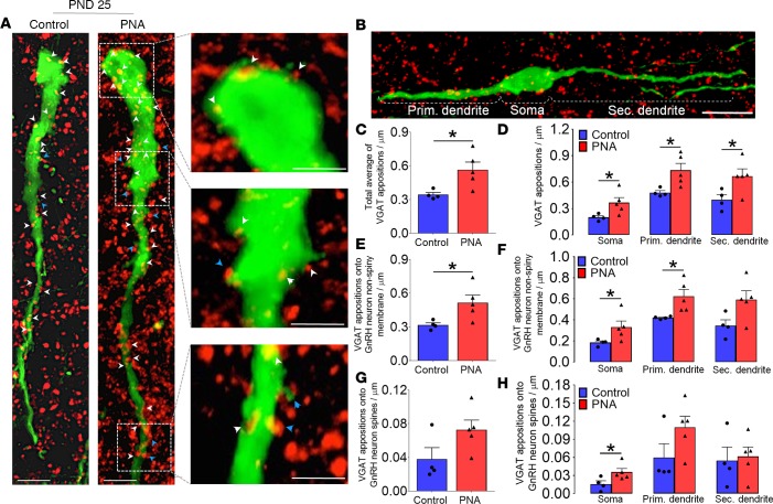
Figure 2. Increased GABAergic contact to GnRH neurons is present in prepubertal PNA mice.
(A) Confocal images of control (N = 4; 45 neurons total) and prenatally androgenized (PNA) (N = 5; 51 neurons total) mice showing GnRH-GFP neurons (green) and vesicular GABA transporter–immunoreactive (VGAT-ir) puncta (red) in the rostral preoptic area. Scale bars: 10 μm. Inset images depict a selected confocal image stack of 1.15 μm thickness illustrating VGAT appositions at the level of GnRH neuron soma and dendrite. Red puncta (VGAT-ir) in close apposition to green GnRH neurons can appear as yellow or with a yellow halo as a result of overlap in confocal projections. White arrowheads point to putative GABA inputs onto the non-spiny GnRH neuron membrane and blue arrowheads show GABA inputs onto spines. Scale bars: 5 μm. (B) Neuronal compartments of bipolar GnRH neurons. Morphological criteria classified the primary dendrite as the thicker dendrite (with largest sectional area leaving the soma) and secondary dendrite as the thinner dendrite. Histograms depict total VGAT-ir apposition density on GnRH neurons (C) and per neuronal compartment (D). Total VGAT-ir apposition density onto the non-spiny GnRH neuron membrane (E), and per neuronal compartment (F). Total VGAT-ir apposition density onto GnRH neuron spines (G), and per neuronal compartment (H). Histogram values are represented as mean ± SEM with dot plot of individual values. *P < 0.05; Mann-Whitney U test. GnRH, gonadotropin-releasing hormone.

