Figure 5.
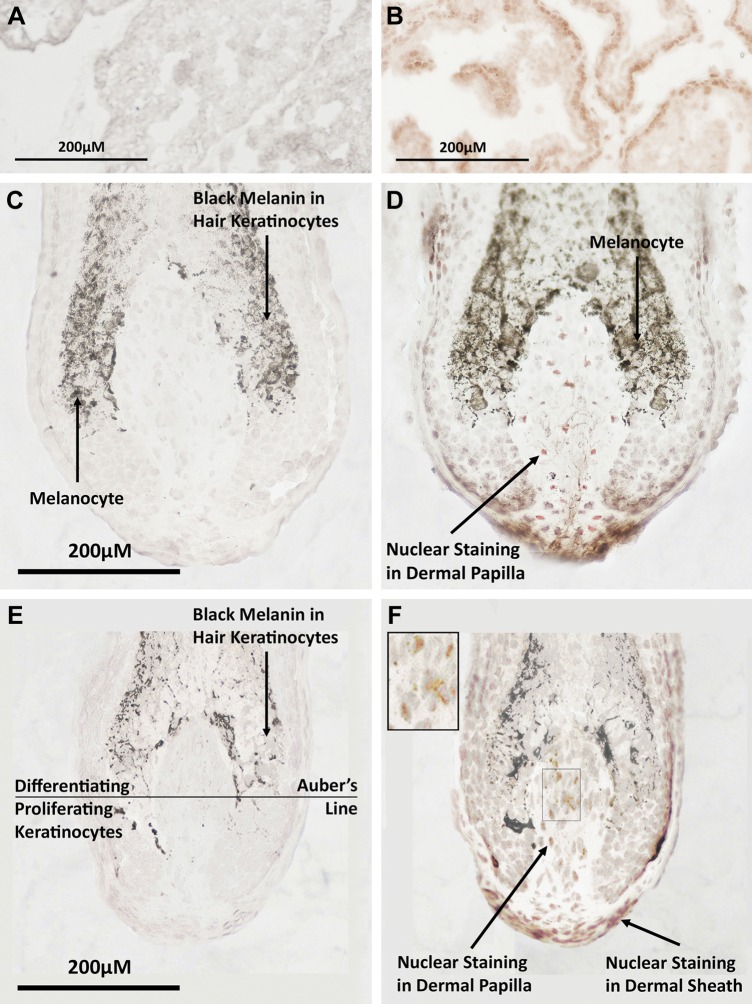
Androgen receptors are present in the hair bulbs of female preauricular hair follicles. A, B) Photomicrographs of frozen sections of positive control adult rat prostate incubated with an mAb to the androgen receptor showed red positive staining in the nuclei of columnar epithelial cells (A), but no staining in the negative control (B). D, F) Immunohistochemistry of human preauricular hair follicle cryosections cut longitudinally through the approximate follicle midpoint located androgen receptors in the nuclei of dermal papilla cells and dermal sheath cells in both terminal (D) and intermediate (F) hair follicles. High-power insert in panel F is at ×2 higher magnification. No staining was observed in keratinocytes in the hair bulbs of either type of follicle. Keratinocytes in the hair bulb below the representative Auber’s line are predominantly dividing, whereas those above are differentiating (52). C, E) Dark brown areas in follicle sections are naturally occurring melanin within hair keratinocytes or melanocytes in the bulb. Negative control sections showed no background staining in either terminal or intermediate follicles.

